iDea – All-in-One platform
for Dry Eye diagnostic
Quick diagnostic of tear film
iDea is the new instrument for individual analysis of the tear film that allows to carry out a quick and detailed structural research of the tear composition. Research on all
layers (Lipid, Aqueous, Mucin) and Meibomian Glands.
Identification of dry eye type
Thanks to the new device it is possible to identify the type of Dry Eye Disease (DED) and determine which components can be treated with a specific treatment, in relation to the type of deficiency.
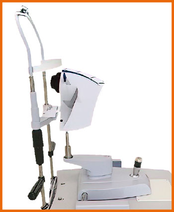
Testing options with iDea
INTERFEROMETRY
iDea automatically evaluates quantity and quality of the lipid component on the tear film. iDea highlights the lipid layer and software analyses automatically Lipid Layer Thickness (LLT).
TEAR MENISCUS
The tear meniscus thickness is observed on the eyelid margins providing useful information on the tear volume. The tear meniscus can be examined considering its height, regularity and shape.
NIBUT
The stability of the mucin layer and the whole tear film is assessed through studying of non-invasive break up time (NIBUT), by using the placido cone projected onto the cornea.
MEIBOGRAPHY
Visualization of glands with meibography through illumination of the eyelid with infrared light. It images the morphology of the glands in order to diagnose any meibomian gland drop out which would lead to tear dysfunction.
BLEPHARITIS
This test helps to visually see blepharitis and presence of demodex. It can be performed on the outer surface of the eye and eyelids.
OCULAR REDNESS CLASSIFICATION
Once the image of the conjunctiva with its blood vessels is captured, it is possible to compare it with the classification sheets of bulbar and limbal redness degrees.
PUPILLOMETRY
Measurement of the pupil reaction to light with and without glare. Measurement mode: scotopic, mesopic, photopic
WHITE TO WHITE MEASUREMENT
Evaluation of corneal diameter from limbus to limbus (white-to-white distance, WTW).
Auto Interferometry
With iDea, interferometry is simple, fast and automatic. Software automatically detects the stained lipids on the patient‘s eye and determines the lipid layer thickness (LLT) based on Guillon‘s international OD study.*
Automatic measurement of:
- Maximum lipid layer thickness
- Average thickness
- Blinking rate
The values are displayed on a user-friendly grading scale to explain the disease to the patient (image 1).
Comparison between meibomian glands and lipid layer thickness to capture the functionality of the meibomian glands before and after treatment (image 2).
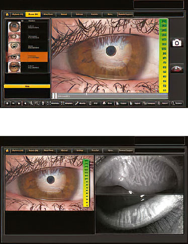
At a glance: Tests with iDea quick diagnostic
- 3D Meibography
- Auto-NIBUT: Non Invasive Break Up Time
- BUT: Break Up Time
- LLT: Lipid Layer Thickness
- T. M.: Tear meniscus
- OSDI: Ocular Surface Disease Index
- Osmolarity
- Schirmer test
- Blink rate
- White-to-white measurements
* Guillon JP. Non-invasive Tearscope Plus routine for contact lens fitting. Cont. Lens Ant. Eye. 1998
Please do not hesitate to contact us for more information.
We would be glad to advise you.


